X-ray Analysis: HIO Series
I wanted to give a little insight into a very important part of our care here at Ohio Specific Chiropractic: the X-ray Analysis.
On each visit, I am trying to answer three simple questions: Where is the vertebral subluxation? When is it a vertebral subluxation? and How can I correct the vertebral subluxation? To answer the Where question, I look to X-ray analysis.
X-rays or Spinographs of the cervical spine help lay down the foundation and blueprint for the patient's individual care. Precision X-rays are taken to establish the exact mal-position of the vertebrae at the location of brainstem interference.
I generally take three specific views of the upper cervical spine and skull to get a 3-dimensional representation of your specific misalignment. X-rays are vital since the tiny and intricate misalignments of the upper cervical spine can’t always be detected by other means such as palpation. We measure the misalignment in millimeters and degrees, so specificity is crucial.
With this analysis, our main point of reference with regards to the misalignment is the foramen magnum. The foramen magnum is the portion of the skull where the brainstem exits and resides within the two top bones of the neck, the Atlas and Axis bones. This aspect of the misalignment can’t be visualized without some form of imaging, such as X-rays.
Lateral View

With this view, we look at the spine from the side and discover the first components of the misalignment. For the Atlas, we determine whether it has gone superior or inferior in relation to the foramen magnum. For the Axis bone, we determine if it has misaligned posterior and/or inferior in relation to the foramen magnum.
Anterior-Posterior Open Mouth View

With the Anterior-Posterior Open Mouth, or APOM for short, we look at the spine from the back to front and discover the next components of the misalignment. For Atlas, we determine whether it has laterality shifted left or right in relation to the foramen magnum. For Axis, we determine if it has rotated and/or laterality shifted left or right in relation to the foramen magnum.
Base Posterior
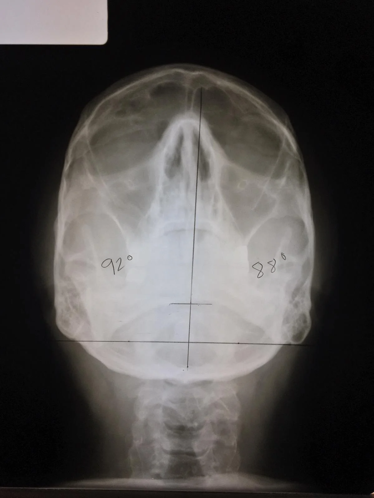
With the Base Posterior, or BP for short, we look at the spine from superior to inferior (top to bottom) in order to discover the final components of the misalignment. For Atlas, we determine whether it has rotated anterior or posterior in relation to the foramen magnum. For the Axis on this view, we use the film to help confirm the previous findings from the other two films.
With our analysis of these 3 views, we can answer “Where is the vertebral subluxation?” We put this together with the other important information gathered from the exam to start the process of tailor-making your adjustment specific for you.
- Jarek Esarco, DC, CACCP
Related Blogs:
Answering Dr. Robert Mendelsohn's X-ray Questions
What to Expect: 9 Steps of Your First Visit at OHIO Specific Chiropractic