100+ Years of Pediatric Spinography
Chiropractors for over 100 years have trained to take and analyze X-Ray films. X-rays are also known as radiographs or by the more common Chiropractic term, spinographs. Spinographs aid in vertebral subluxation detection.
Chiropractors have over 300 hours of radiography education. This includes X-ray analysis, equipment use and patient placement. The study of spinography accounts for 12% of our clinical training in college.
X-rays were first discovered in 1895 by physicist Wilhelm Roentgen. The first known X-ray film was taken of his wife’s hand, Anna Roentgen, that same year. Medicine early on saw the potential to implement X-rays as part of their diagnosis procedures.

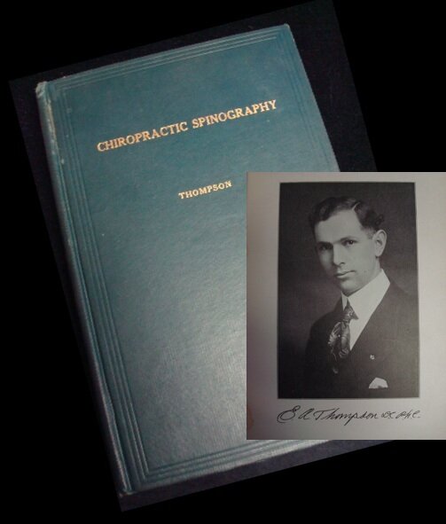
Chiropractic, also discovered in 1895, was not far behind to incorporate X-rays into practice as well. Dr. B.J. Palmer first added X-rays to the curriculum in 1910 after extensive research. He saw great potential for X-rays to improve the analysis of determining the exact position of the vertebral subluxation. Dr. Enerst Thompson took B.J. Palmer’s work to print in his 1918 published textbook, Chiropractic Spinography. Quoting Dr. Thompson:
“The X-ray has become one of the most valuable clinical examination tools available. The graphical output of anatomical structures of the human body produced by X-rays generally serves as a “true picture” of the X-ray object.”
Up to this point, the only way to determine the misalignment of the subluxation was by palpation and nerve tracing. But palpation and nerve tracing is fraught with inconsistencies and subjectivity. With palpation, you can only feel the outer structures of the spine, the spinous process and the transverse processes. But these structures are often asymmetrical or bent and can provide false readings. Nowhere are these discrepancies noticed more than the upper cervical spine, especially with the Atlas bone. Quoting Dr. Palmer:
“It must be freely admitted that this particular vertebra [Atlas], above all others, is the most difficult to palpate and requires the spinograph more often to determine the true condition.”

If we look to the textbook definition of a vertebral subluxation, it has four components:
Loss of juxtaposition to the vertebra above, below or both
Occlusion of a neural foramen
Compromised neural integrity or function
Interference to the flow of mental impulses
The first two components regard the skeletal system. The third and fourth concern the nerve system. It is possible to determine the loss of juxtaposition by palpation. But again, not always reliable or verifiable through palpation alone. You can not determine neural foramen occlusion without some form of imaging.
There are two major types of neural foramen found in the spine. They are the vertebral foramen and the intervertebral foramen. A vertebral foramen is the opening found on each spinal bone that encompasses the brainstem or spinal cord.

An intervertebral foramen is created between two spinal bones and is how a spinal nerve exits the spinal cord. You can not palpate either of these internal structures. X-rays help determine the size and shape of both foramina. To be certain of a vertebral subluxation, it is imperative to be able to “check off all the boxes” that apply to its definition.
The importance of determining foramen involvement applies to everyone, no matter their age. Everyone is susceptible to nerve disruption from a subluxation. Quoting again from Dr. Palmer:
“The spinograph always discloses the true condition and its use is always advisable when the adjuster is not positive of the subluxation.”
Now, when I say that “everyone is susceptible to nerve disruption,” do I mean everyone? Even infants and children? Absolutely! Dr. John H. Craven wrote about this idea in his 1924 textbook Chiropractic Hygiene and Pediatrics. Quoting:
“Whenever possible a spinograph should be made of the child...Every possible means should be used to verify the palpation.”

The pediatric population is more susceptible to nerve disruption because of their impressionable quality. The spine of a developing child is influenced greatly by the stresses placed on it. A spine needs only to misalign a few millimeters to compromise nerve integrity. The same concentration to detail and care is paramount in this special demographic to pinpoint these minute changes in biomechanics. X-rays help with this matter.

While X-rays do have great potential to assist in the analysis of a vertebral subluxation, they are not without potential risk. Radiation exposure is a concern, but was addressed early on in the profession. Quoting again from Dr. Thompson:
“Extreme care must be exercised in taking spinographs of children, or they will be absolutely useless, because of the danger of over-exposure or of the child moving during the exposure.”
The process of obtaining an X-ray image demands attention to safety and is always on the mind of the Chiropractor. There is an old Chiropractic adage that goes along these lines:
Why X-rays? To see is to know. To not see is to guess. And we don't guess when it comes to your health.
Speculation can place you on shaky ground. As a Chiropractor, I want to make sure my care is set on a solid base. X-rays help give strength and stability to the foundation of my care, the adjustment.
- Jarek Esarco, DC, CACCP
Related Blogs:
Adjusting Children: A New Age Niche or an Established Institute Foundation?
Answering Dr. Robert Mendelsohn’s X-ray Questions
Can Infants Get Vertebral Subluxations?
Top 3 Reasons Why Parents Take Their Children to a Family Wellness Chiropractor
Safety and Effectiveness of Pediatric Chiropractic